Difference between revisions of "Traumatic Brain Injury"
Techsensus (talk | contribs) |
Techsensus (talk | contribs) (→State of the Art) |
||
| (15 intermediate revisions by the same user not shown) | |||
| Line 29: | Line 29: | ||
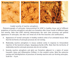
Very important cell types in the brain, aside from neurons, are the neuroglia. These cell types are broadly recognized as having a supportive role. There are 6 types of neuroglia, each with their own function [[File:Types-of-neuroglia_brain-physiology-cells-QBI.png|thumb|250px|Types of glia. <ref name="Arti16">https://qbi.uq.edu.au/brain-basics/brain/brain-physiology/types-glia</ref>]]. In the central nervous system (CNS), the astrocytes are a main type of glial cells. They contribute to the maintenance of homeostasis in the CNS through reactive astrogliosis.<ref name="Arti14">Sofroniew, M. V., & Vinters, H. V. (2009). Astrocytes: biology and pathology. Acta Neuropathologica, 119(1), 7–35. https://doi.org/10.1007/s00401-009-0619-8 </ref> This process is triggered by all types of brain insults and has features that aid recovery, but simultaneously have a potential to inflict damage on the brain. For example, the astrocytes break down their glycogen to supply adjacent neurons with lactate, which the neurons use as fuel to recover. However, the astrocytes also play a critical role in water movements through the brain and in pathological conditions, this can mediate oedema. In all cases of reactive astrogliosis, GFAP is up-regulated. The degree to which the GFAP expression changes, is only dependent on the severity of the trauma to the CNS and not on the morphological appearance of the reactive astrogliosis. <ref name="Arti15">Verkhratsky, A., & Butt, A. (2013). General Pathophysiology of Neuroglia. In Glial Physiology and Pathophysiology (1st ed.). John Wiley & Sons, Ltd. https://doi.org/10.1002/9781118402061 </ref> | Very important cell types in the brain, aside from neurons, are the neuroglia. These cell types are broadly recognized as having a supportive role. There are 6 types of neuroglia, each with their own function [[File:Types-of-neuroglia_brain-physiology-cells-QBI.png|thumb|250px|Types of glia. <ref name="Arti16">https://qbi.uq.edu.au/brain-basics/brain/brain-physiology/types-glia</ref>]]. In the central nervous system (CNS), the astrocytes are a main type of glial cells. They contribute to the maintenance of homeostasis in the CNS through reactive astrogliosis.<ref name="Arti14">Sofroniew, M. V., & Vinters, H. V. (2009). Astrocytes: biology and pathology. Acta Neuropathologica, 119(1), 7–35. https://doi.org/10.1007/s00401-009-0619-8 </ref> This process is triggered by all types of brain insults and has features that aid recovery, but simultaneously have a potential to inflict damage on the brain. For example, the astrocytes break down their glycogen to supply adjacent neurons with lactate, which the neurons use as fuel to recover. However, the astrocytes also play a critical role in water movements through the brain and in pathological conditions, this can mediate oedema. In all cases of reactive astrogliosis, GFAP is up-regulated. The degree to which the GFAP expression changes, is only dependent on the severity of the trauma to the CNS and not on the morphological appearance of the reactive astrogliosis. <ref name="Arti15">Verkhratsky, A., & Butt, A. (2013). General Pathophysiology of Neuroglia. In Glial Physiology and Pathophysiology (1st ed.). John Wiley & Sons, Ltd. https://doi.org/10.1002/9781118402061 </ref> | ||
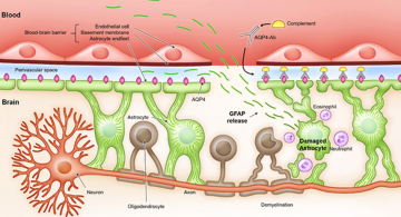
| − | When the cause of the increase in GFAP production is brain injury, one can imagine there are several ways for the molecule to end up in the extracellular space instead of remaining in the intracellular space where it was produced [[File:Expression of GFAP in reactive astrogliosis.png|thumb| | + | When the cause of the increase in GFAP production is brain injury, one can imagine there are several ways for the molecule to end up in the extracellular space instead of remaining in the intracellular space where it was produced [[File:Expression of GFAP in reactive astrogliosis.png|thumb|270px|Expression of GFAP in reactive astrogliosis.<ref name="Arti18">Verkhratsky, A., & Butt, A. (2013). General Pathophysiology of Neuroglia. In Glial Physiology and Pathophysiology (1st ed.). John Wiley & Sons, Ltd. https://doi.org/10.1002/9781118402061</ref>]]. To give an example, necrosis leads to leakage of cellular molecules.<ref name="Arti19">Messing, A., & Brenner, M. (2020). GFAP at 50. ASN Neuro, 12, 175909142094968. https://doi.org/10.1177/1759091420949680</ref> Due to the greater permeability of the BBB, GFAP concentrations increase in the blood as well. Within the first hour of injury, elevated levels of serum GFAP can be detected. At twenty hours of injury, the serum level of GFAP reaches its peak. During the next 52 hours, the GFAP level will decrease slowly. Moreover, there is no significant increase in serum levels of GFAP in patients without TBI.<ref name="Arti17">Abdelhak, A., Foschi, M., Abu-Rumeileh, S., Yue, J. K., D’Anna, L., Huss, A., Oeckl, P., Ludolph, A. C., Kuhle, J., Petzold, A., Manley, G. T., Green, A. J., Otto, M., & Tumani, H. (2022). Blood GFAP as an emerging biomarker in brain and spinal cord disorders. Nature Reviews Neurology, 18(3), 158–172. https://doi.org/10.1038/s41582-021-00616-3</ref> Therefore, a biosensor that detects the levels of GFAP can be used to determine the presence of traumatic brain injury in patients who have received a significant force to their head. |
| + | |||
| + | == Medical application and relevance == | ||
| + | The global incidence of TBI is estimated to be 27 to 69 million a year <ref name="Arti20">Dewan, M. C., Rattani, A., Gupta, S., Baticulon, R. E., Hung, Y. C., Punchak, M., Agrawal, A., Adeleye, A. O., Shrime, M. G., Rubiano, A. M., Rosenfeld, J. V., & Park, K. B. (2019). Estimating the global incidence of traumatic brain injury. Journal of Neurosurgery, 130(4), 1080–1097. https://doi.org/10.3171/2017.10.jns17352</ref> <ref name="Arti21">James, S. L., Theadom, A., Ellenbogen, R. G., Bannick, M. S., Montjoy-Venning, W., Lucchesi, L. R., Abbasi, N., Abdulkader, R., Abraha, H. N., Adsuar, J. C., Afarideh, M., Agrawal, S., Ahmadi, A., Ahmed, M. B., Aichour, A. N., Aichour, I., Aichour, M. T. E., Akinyemi, R. O., Akseer, N., . . . Murray, C. J. L. (2019). Global, regional, and national burden of traumatic brain injury and spinal cord injury, 1990–2016: a systematic analysis for the Global Burden of Disease Study 2016. The Lancet Neurology, 18(1), 56–87. https://doi.org/10.1016/s1474-4422(18)30415-0</ref>. Across all severities of TBI, mortality is quite low at 3% <ref name="Arti22">Georges, A., & Das, J. M. (2022). Traumatic Brain Injury [Internet]. In StatPearls. StatPearls Publishing. https://www.ncbi.nlm.nih.gov/books/NBK459300/</ref> this was determined in the United States of America, a country where the healthcare system is quite developed. While mortality is low, the long term effects of TBI can be detrimental to a person’s quality of life. Some of the acute symptoms lessen or resolve over time, such as dizziness or nausea. Other consequences do not become apparent until a long period of time has passed, for instance psychiatric conditions.<ref name="Arti23">Seel, R. T., Macciocchi, S., & Kreutzer, J. S. (2010). Clinical Considerations for the Diagnosis of Major Depression After Moderate to Severe TBI. Journal of Head Trauma Rehabilitation, 25(2), 99–112. https://doi.org/10.1097/htr.0b013e3181ce3966</ref> | ||
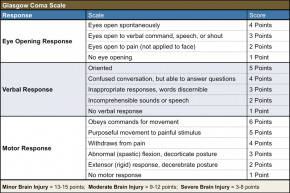
| + | Currently, the Glasgow coma scale (GCS) is the only standardized way to assess patients with a suspected TBI. The GCS provides a practical method for assessing impairment of conscious level in response to defined stimuli.[[File:The glasgow coma scale.png|thumb|290px|The Glasgow coma scale.<ref name="Arti24">https://smhs.gwu.edu/urgentmatters/news/keep-it-simple-acute-gcs-score-binary-decision</ref>]] Depending on the final score, the TBI can be classified as minor, moderate or severe. | ||
| + | |||
| + | While the GCS score describes the current condition of the patient, its usage has many shortcomings, one of which is the fact that physicians seem to struggle with remembering the exact levels of the scale.<ref name="Arti25">Riechers, R. G., Ramage, A., Brown, W., Kalehua, A., Rhee, P., Ecklund, J. M., & Ling, G. S. (2005). Physician Knowledge of the Glasgow Coma Scale. Journal of Neurotrauma, 22(11), 1327–1334. https://doi.org/10.1089/neu.2005.22.1327</ref> Although there is a dose-response relationship with regard to the severity of the TBI and the severity of the outcome, the GCS does not effectively and uniformly predict mortality rate. <ref name="Arti26">Institute of Medicine (US) Committee on Gulf War and Health: Brain Injury in Veterans and Long-Term Health Outcomes. (2008). Gulf War and Health: Volume 7 (Vol. 7) [Internet]. National Academies Press. https://doi.org/10.17226/12436</ref> <ref name="Arti27">Cho, D. Y., & Wang, Y. C. (1997). Comparison of the APACHE III, APACHE II and Glasgow Coma Scale in acute head injury for prediction of mortality and functional outcome. Intensive Care Medicine, 23(1), 77–84. https://doi.org/10.1007/s001340050294</ref> | ||
| + | |||
| + | Other ways for diagnosing TBI can include various imaging tests like a CT scan or an MRI scan. However, these imaging techniques are often not sensitive/specific enough for milder TBI. According to experts, only 5-10% of mild TBI patients have abnormal CT/MRI scans. The problem with these scans is that they can only detect damage on a macroscopic level, whereas mild TBI manifests primarily at a microscopic level. Therefore, doctors can mistakenly believe that patients with a standard CT or MRI scan have not suffered a TBI.<ref name="Arti28">McKinlay, A., Lin, A., & Than, M. (2018). A comparison of emergency department medical records to parental self-reporting of traumatic brain injury symptoms. Concussion, 3(1), CNC52. https://doi.org/10.2217/cnc-2017-0017</ref> All in all, improvements are needed to objectively and effectively determine whether or not a patient is suffering from a TBI. | ||
| + | |||
| + | == State of the Art == | ||
| + | Currently, the concentration of GFAP is typically measured in blood plasma (liquid component of blood in which blood cells are absent) or serum (plasma from which the clotting proteins have been removed). Classic methods for the detection of GFAP include ELISA (Enzyme-Linked Immunosorbent Assay). | ||
| + | |||
| + | {| class="wikitable" style="margin-bottom:0" | ||
| + | !Test Name | ||
| + | !Sample Volume (μL) | ||
| + | !Sample matrix | ||
| + | !Range (pg/mL) | ||
| + | !Precision (CV%) | ||
| + | !Incubation time (min) | ||
| + | !Measuring Technique | ||
| + | !Assay type | ||
| + | !Links | ||
| + | |- | ||
| + | |GFAP CLIA Assay, from Eagle Biosciences | ||
| + | |100 | ||
| + | |serum, CSF | ||
| + | |10-640 | ||
| + | |Intra-Assay: CV=1%, Inter-Assay: CV=6% | ||
| + | |150 | ||
| + | |ELISA | ||
| + | |solid phase enzyme-linked sandwich immunosorbent | ||
| + | |https://eaglebio.com/product/gfap-clia-assay-kit/ https://eaglebio.com/wp-content/uploads/2022/01/gfp31-L01-gfap-clia-assay-kit-package-insert-2022.pdf | ||
| + | |- | ||
| + | |Human GFAP ELISA Kit, from FineTest | ||
| + | | | ||
| + | |serum, plasma, tissue homogenates, other biological fluids | ||
| + | |313-20000 | ||
| + | |Intra-Assay: CV<8%, Inter-Assay: CV<10% | ||
| + | | | ||
| + | |ELISA | ||
| + | |sandwich | ||
| + | |https://www.fn-test.com/product/eh0410/ https://www.fn-test.com/content/uploads/product/manuals/elisa/EH0410.pdf | ||
| + | |- | ||
| + | |Human GFAP ELISA Kit, Producer not known, distributed by Mybiosource | ||
| + | |>10 | ||
| + | |serum, plasma, tissue homogenates | ||
| + | |625-40000 | ||
| + | |Intra-Assay: CV<8%, Inter-Assay: CV<10% | ||
| + | |≈300 | ||
| + | |ELISA | ||
| + | |quantitative sandwich enzyme immunoassay | ||
| + | |https://www.mybiosource.com/gfap-human-elisa-kits/glial-fibrillary-acidic-protein/704044 https://cdn.mybiosource.com/tds/protocol_manuals/000000-799999/MBS704044.pdf | ||
| + | |- | ||
| + | |Human GFAP ELISA Kit, from LSBio | ||
| + | |>10 | ||
| + | |plasma, serum | ||
| + | |156 - 10000 | ||
| + | |Intra-Assay: CV<10%, Inter-Assay: CV<12% | ||
| + | |≈300 | ||
| + | |ELISA | ||
| + | |sandwich | ||
| + | |https://www.lsbio.com/elisakits/human-gfap-sandwich-elisa-elisa-kit-ls-f4258/4258?trid=247 https://www.lsbio.com/elisakits/manualpdf/ls-f4258.pdf | ||
| + | |- | ||
| + | |GFAP Human ProcartaPlex™ Simplex Kit, from Thermo | ||
| + | |>25 | ||
| + | |plasma, serum, CSF | ||
| + | |8 - 32900 | ||
| + | | | ||
| + | | | ||
| + | |ELISA | ||
| + | |sandwich | ||
| + | |https://www.thermofisher.com/order/catalog/product/EPX010-12336-901 | ||
| + | |- | ||
| + | |GFAP ELISA Kit, from Aviva Systems Biology | ||
| + | |>2 | ||
| + | |Serum, plasma, other biological fluids | ||
| + | |31.25-2000 | ||
| + | |Intra-Assay: CV<10%, Inter-Assay: CV<12% | ||
| + | |300 | ||
| + | |ELISA | ||
| + | |sandwich enzyme-linked immuno-sorbent | ||
| + | |https://www.avivasysbio.com/gfap-elisa-kit-bovine-okcd02567.html https://www.avivasysbio.com/pub/media/pdf/products/OKCD02567.pdf | ||
| + | |- | ||
| + | |}<div style="margin-bottom:1em"><sub>''Table 1: Commercially available GFAP ELISA test assays.</sub><br/> | ||
| + | |||
| + | As is clear from the above table, the classic ELISA technique is widely available as commercial kits, which makes it a convenient method for biomarker detection in patient samples. Nevertheless, a relatively long detection time (of at least a few hours) and the requirement of a laboratory environment precludes its use in GP surgeries or patients’ homes.<ref name="Arti29">Xu, L., Ramadan, S., Akingbade, O. E., Zhang, Y., Alodan, S., Graham, N., Zimmerman, K. A., Torres, E., Heslegrave, A., Petrov, P. K., Zetterberg, H., Sharp, D. J., Klein, N., & Li, B. (2021). Detection of Glial Fibrillary Acidic Protein in Patient Plasma Using On-Chip Graphene Field-Effect Biosensors, in Comparison with ELISA and Single-Molecule Array. ACS Sensors, 7(1), 253–262. https://doi.org/10.1021/acssensors.1c02232</ref> Classic ELISAs are laboratory-based assay, which are not suited for rapid testing as targeted in the SensUs competition.The same applies to more advanced laboratory assays, such as mass spectrometry and single molecular array (Simoa) technology. | ||
| + | Recently a commercial GFAP test has been developed by Abbott on its i-STAT Alinity biosensor platform. The specifications are listed in the Table below. The i-STAT platform is used in hospitals. However, the i-STAT TBI Plasma test is not intended to be used in point-of-care settings. | ||
| + | |||
| + | {| class="wikitable" style="margin-bottom:0" | ||
| + | !Test Name | ||
| + | !Sample Volume (μL) | ||
| + | !Sample matrix | ||
| + | !Range (pg/mL) | ||
| + | !Precision (CV%) | ||
| + | !Incubation time (min) | ||
| + | !Measuring Technique | ||
| + | !Assay type | ||
| + | !Links | ||
| + | |- | ||
| + | |i-STAT TBI Plasma | ||
| + | |>20 | ||
| + | |plasma | ||
| + | |30-10000 | ||
| + | | | ||
| + | |15 | ||
| + | |cartridge for analyzer | ||
| + | | | ||
| + | |https://www.globalpointofcare.abbott/en/product-details/apoc/istat-tbi-plasma.html https://www.globalpointofcare.abbott/en/product-details/apoc/istat-alinity.html | ||
| + | |- | ||
| + | |}<div style="margin-bottom:1em"><sub>''Table 2: Commercially available GFAP biosensor.</sub><br/> | ||
| + | |||
| + | == Lab protocols == | ||
| + | During the preparation of the blood samples, one should avoid physical contact with blood by wearing gloves, glasses, and lab coats. Needles and lancets should be used only once and disposed of in a sharps container for decontamination. Furthermore, cuts already present on hands or arms should be covered with plasters to avoid blood-on-blood contact. After completing the samples, gloves should be removed and hands should be washed thoroughly. The waste should be disposed of in specific biohazard waste bins or bags. | ||
| + | |||
== References == | == References == | ||
<references /> | <references /> | ||
Latest revision as of 10:05, 22 December 2022

Contents
General information
The theme of SensUs 2023 is Traumatic Brain Injury (TBI). It is often stated in the literature that TBI is a silent epidemic with an estimated 64–74 million new cases presenting each year.[2] The impairments suffered by many TBI patients, such as memory loss, cognitive dysfunction, or behavioural disturbance, are often not visible. The economic and social impact is considerable, with an estimate of direct medical expenditures and indirect costs (e.g., loss of productivity) attributed to TBI exceeding $60 billion in 2000 in the USA. TBI is defined as an alteration in brain function, or other evidence of brain pathology, caused by an external force.[3] The severity of injury in TBI is classified as mild, moderate, or severe with mild TBIs being the most common.[4] Alteration in brain function can be manifest by loss or decreased level of consciousness, alteration in mental state, incomplete memory for the event, or neurological deficits. Examples of external forces include the head striking or being struck by an object, rapid acceleration or deceleration of the brain, penetration of the brain by a foreign object, and exposure to forces associated with blasts.
GFAP is a protein found in the glial cells of neural tissue. Glial cells are non-neuronal cells that provide physical and metabolic support to neurons. Blood biomarker levels of GFAP reflect acute injury to brain tissue since the biomarker levels increase strongly and fast within hours after the trauma occurred. Therefore, it has potential use in the emergency unit and intensive care unit, directly after the TBI. Furthermore, the biomarker persists for an extended period with a half-life of 48 hours, making it a favourable biomarker to use in both the acute and subacute phases of injury. The concentration of GFAP peaks at about 20 hours after the injury. Reference serum levels of GFAP range from 0.02 — 0.35 𝜇g/L and the cut-off value of a negative CT scan for GFAP is 0.35 𝜇g/L.[5]
History of TBI
Pre-historic period
TBIs play an important role in the evolution of humans. Studies of skeletons from the mesolithic period (c. 7000 BC) suggest an inverse relationship between the risk of skull fracture and the progress of civilization. The palaeolithic period is a period in prehistory that occurred over a million years ago. In those times, man's predecessor was a semi-erect hominid now named Australopithecus africanus; a damaged skull from this species found in South Africa reveals the first evidence of brain injury.[6]
One of the earliest methods used for treating TBI is called Trepanation. Trepanning, also known as trepanation, is a surgical intervention in which a hole is drilled or scraped into the human skull. The earliest evidence of trepanation performed by the man himself appears in the Neolithic period. The primary theories for the practice of trepanation in ancient times include spiritual purposes and treatment for epilepsy, headache, head wound, and mental disorders.
Ancient History
The first written evidence of TBI is documented in an ancient Egyptian text known as the Edwin Smith Papyrus which was written around 1650-1550 BC and believed to be a copy of an even older manuscript written around 3000 BC. It describes various head injuries and symptoms and classifies them based on their presentation and tractability.[7] This text is so old that it goes as far as mentioning magic as a last resort in terminal cases. To get an idea of what treatment for a brain injury was like then, bandaging the head wound with meat and applying a honey and oil type of dressing until healed were common methods.[8] There are several biblical references to head injuries which are reported to have occurred in the 12-10th century BC. These references to head injuries include: Sisera’s death at the hands of Jael, skull fractures on Abimelech, and the most famous death of Goliath by David.[9]
Hippocrates of Kos, also known as Hippocrates II, was a Greek physician of the classical period who is considered one of the most outstanding figures in the history of medicine and is referred to as the "Father of Medicine”. The first systematic approach to head injuries was by Hippocrates. The Hippocratic Corpus consisted of 76 Treatises, one of which was "On Injuries of the Head".[10]
Medieval History
In the Middle Ages, physicians further described head injury symptoms and the term concussion became more widespread. Berengario da Carpi, an Italian physician, published one of the first books on head injuries. He categorized head injuries into lacerations, contusions and perforations, each of which could be associated with a fracture. A prime example from the 16th century is also one of history’s most well-known figures. A recent study argues that King Henry VIII of England’s erratic behavior was a result of possible repeated traumatic brain injuries. Researchers have made a compelling case citing notes that describe changes including memory loss, irritability, impulsive nature, and insomnia. All known today as common symptoms of a traumatic brain injury.[11]
Modern Era
It was first suggested in the 18th century that symptoms arising from a head injury are not due to a fractured skull, but to injury of the brain. Percival Pott (1713-1788) was one of the first to emphasize that it was the neurological status and not the skull fracture that determined whether surgical intervention was indicated. One of the most notable instances of brain injury took place in the 1800s. Phineas Gage survived an accident where an iron rod penetrated his head and destroyed a good portion of his left frontal lobe. Prior to his accident, Gage was described as “even-tempered”, but his demeanor shifted significantly afterwards. Due to the definitive nature of his injury and the personality changes that followed, many cite this as the first case illustrating mood and personality shifts directly resulting from a brain injury. The 20th century saw the advancement of technologies that improved treatment and diagnosis such as the development of imaging tools including CT and MRI, and, in the 21st century, diffusion tensor imaging (DTI). The introduction of intracranial pressure monitoring in the 1950s has been credited with beginning the "modern era" of head injury.[12] In the 1970s, awareness of TBI as a public health problem grew, and a great deal of progress has been made since then in brain trauma research, such as the discovery of primary and secondary brain injury. Prevention of TBI has also become immensely important with several efforts made in that direction such as: introduction of helmets in the army and motorcyclists, airbags in motor vehicles etc. The 1990s saw the development and dissemination of standardized guidelines for the treatment of TBI, with protocols for a range of issues such as drugs and management of intracranial pressure.
Mechanism of Traumatic Brain Injury
The mechanical force applied to the head displaces the brain. Depending on the direction and magnitude of the force, certain neurological functions can be disrupted. The damage to the brain within the skull can present as strain, tissue distortion and shearing of axons. The processes for damage control are activated immediately, while the damage continues to evolve. The damage occurs as a result of the cellular and molecular processes taking place in response to mechanical forces. First, the mechanical force disrupts the neuronal membrane, which rapidly increases the extracellular potassium concentration. This activates a positive feedback loop where more potassium ions are released, which leads to a cascade of responses that eventually lead to a period of hyperglycolysis in the brain followed by higher permeability of the Blood-Brain-Barrier (BBB) and the induction of cytotoxic edema in the brain.[13] The latter has a detrimental effect on all cells in the brain tissue.
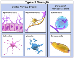
Very important cell types in the brain, aside from neurons, are the neuroglia. These cell types are broadly recognized as having a supportive role. There are 6 types of neuroglia, each with their own function

Medical application and relevance
The global incidence of TBI is estimated to be 27 to 69 million a year [20] [21]. Across all severities of TBI, mortality is quite low at 3% [22] this was determined in the United States of America, a country where the healthcare system is quite developed. While mortality is low, the long term effects of TBI can be detrimental to a person’s quality of life. Some of the acute symptoms lessen or resolve over time, such as dizziness or nausea. Other consequences do not become apparent until a long period of time has passed, for instance psychiatric conditions.[23]
Currently, the Glasgow coma scale (GCS) is the only standardized way to assess patients with a suspected TBI. The GCS provides a practical method for assessing impairment of conscious level in response to defined stimuli.
While the GCS score describes the current condition of the patient, its usage has many shortcomings, one of which is the fact that physicians seem to struggle with remembering the exact levels of the scale.[25] Although there is a dose-response relationship with regard to the severity of the TBI and the severity of the outcome, the GCS does not effectively and uniformly predict mortality rate. [26] [27]
Other ways for diagnosing TBI can include various imaging tests like a CT scan or an MRI scan. However, these imaging techniques are often not sensitive/specific enough for milder TBI. According to experts, only 5-10% of mild TBI patients have abnormal CT/MRI scans. The problem with these scans is that they can only detect damage on a macroscopic level, whereas mild TBI manifests primarily at a microscopic level. Therefore, doctors can mistakenly believe that patients with a standard CT or MRI scan have not suffered a TBI.[28] All in all, improvements are needed to objectively and effectively determine whether or not a patient is suffering from a TBI.
State of the Art
Currently, the concentration of GFAP is typically measured in blood plasma (liquid component of blood in which blood cells are absent) or serum (plasma from which the clotting proteins have been removed). Classic methods for the detection of GFAP include ELISA (Enzyme-Linked Immunosorbent Assay).
| Test Name | Sample Volume (μL) | Sample matrix | Range (pg/mL) | Precision (CV%) | Incubation time (min) | Measuring Technique | Assay type | Links |
|---|---|---|---|---|---|---|---|---|
| GFAP CLIA Assay, from Eagle Biosciences | 100 | serum, CSF | 10-640 | Intra-Assay: CV=1%, Inter-Assay: CV=6% | 150 | ELISA | solid phase enzyme-linked sandwich immunosorbent | https://eaglebio.com/product/gfap-clia-assay-kit/ https://eaglebio.com/wp-content/uploads/2022/01/gfp31-L01-gfap-clia-assay-kit-package-insert-2022.pdf |
| Human GFAP ELISA Kit, from FineTest | serum, plasma, tissue homogenates, other biological fluids | 313-20000 | Intra-Assay: CV<8%, Inter-Assay: CV<10% | ELISA | sandwich | https://www.fn-test.com/product/eh0410/ https://www.fn-test.com/content/uploads/product/manuals/elisa/EH0410.pdf | ||
| Human GFAP ELISA Kit, Producer not known, distributed by Mybiosource | >10 | serum, plasma, tissue homogenates | 625-40000 | Intra-Assay: CV<8%, Inter-Assay: CV<10% | ≈300 | ELISA | quantitative sandwich enzyme immunoassay | https://www.mybiosource.com/gfap-human-elisa-kits/glial-fibrillary-acidic-protein/704044 https://cdn.mybiosource.com/tds/protocol_manuals/000000-799999/MBS704044.pdf |
| Human GFAP ELISA Kit, from LSBio | >10 | plasma, serum | 156 - 10000 | Intra-Assay: CV<10%, Inter-Assay: CV<12% | ≈300 | ELISA | sandwich | https://www.lsbio.com/elisakits/human-gfap-sandwich-elisa-elisa-kit-ls-f4258/4258?trid=247 https://www.lsbio.com/elisakits/manualpdf/ls-f4258.pdf |
| GFAP Human ProcartaPlex™ Simplex Kit, from Thermo | >25 | plasma, serum, CSF | 8 - 32900 | ELISA | sandwich | https://www.thermofisher.com/order/catalog/product/EPX010-12336-901 | ||
| GFAP ELISA Kit, from Aviva Systems Biology | >2 | Serum, plasma, other biological fluids | 31.25-2000 | Intra-Assay: CV<10%, Inter-Assay: CV<12% | 300 | ELISA | sandwich enzyme-linked immuno-sorbent | https://www.avivasysbio.com/gfap-elisa-kit-bovine-okcd02567.html https://www.avivasysbio.com/pub/media/pdf/products/OKCD02567.pdf |
As is clear from the above table, the classic ELISA technique is widely available as commercial kits, which makes it a convenient method for biomarker detection in patient samples. Nevertheless, a relatively long detection time (of at least a few hours) and the requirement of a laboratory environment precludes its use in GP surgeries or patients’ homes.[29] Classic ELISAs are laboratory-based assay, which are not suited for rapid testing as targeted in the SensUs competition.The same applies to more advanced laboratory assays, such as mass spectrometry and single molecular array (Simoa) technology. Recently a commercial GFAP test has been developed by Abbott on its i-STAT Alinity biosensor platform. The specifications are listed in the Table below. The i-STAT platform is used in hospitals. However, the i-STAT TBI Plasma test is not intended to be used in point-of-care settings.
| Test Name | Sample Volume (μL) | Sample matrix | Range (pg/mL) | Precision (CV%) | Incubation time (min) | Measuring Technique | Assay type | Links |
|---|---|---|---|---|---|---|---|---|
| i-STAT TBI Plasma | >20 | plasma | 30-10000 | 15 | cartridge for analyzer | https://www.globalpointofcare.abbott/en/product-details/apoc/istat-tbi-plasma.html https://www.globalpointofcare.abbott/en/product-details/apoc/istat-alinity.html |
Lab protocols
During the preparation of the blood samples, one should avoid physical contact with blood by wearing gloves, glasses, and lab coats. Needles and lancets should be used only once and disposed of in a sharps container for decontamination. Furthermore, cuts already present on hands or arms should be covered with plasters to avoid blood-on-blood contact. After completing the samples, gloves should be removed and hands should be washed thoroughly. The waste should be disposed of in specific biohazard waste bins or bags.
References
- ↑ Glial Fibrillary Acidic Protein in Blood as a Disease Biomarker of Neuromyelitis Optica Spectrum Disorders. Front. Neurol., 17 March 2022, https://www.frontiersin.org/articles/10.3389/fneur.2022.865730/full#F1
- ↑ Rattani, A., Gupta, S., Baticulon, R. E., Hung, Y. C., Punchak, M., Agrawal, A., Adeleye, A. O., Shrime, M. G., Rubiano, A. M., Rosenfeld, J. V., & Park, K. B. (2019). Estimating the global incidence of traumatic brain injury. Journal of Neurosurgery, 130(4), 1080–1097. https://doi.org/10.3171/2017.10.jns17352
- ↑ Menon, D. K., Schwab, K., Wright, D. W., & Maas, A. I. (2010). Position Statement: Definition of Traumatic Brain Injury. Archives of Physical Medicine and Rehabilitation, 91(11), 1637–1640. https://doi.org/10.1016/j.apmr.2010.05.017
- ↑ Iaccarino, C., Gerosa, A., & Viaroli, E. (2021). Epidemiology of Traumatic Brain Injury. In S. Honeybul & A. G. Kolias (Eds.), Traumatic Brain Injury. Springer. https://link.springer.com/chapter/10.1007/978-3-030-78075-3_1
- ↑ Papa, L., Brophy, G. M., Welch, R. D., Lewis, L. M., Braga, C. F., Tan, C. N., Ameli, N. J., Lopez, M. A., Haeussler, C. A., Mendez Giordano, D. I., Silvestri, S., Giordano, P., Weber, K. D., Hill-Pryor, C., & Hack, D. C. (2016). Time Course and Diagnostic Accuracy of Glial and Neuronal Blood Biomarkers GFAP and UCH-L1 in a Large Cohort of Trauma Patients With and Without Mild Traumatic Brain Injury. JAMA Neurology, 73(5), 551. https://doi.org/10.1001/jamaneurol.2016.0039
- ↑ Rose, F. C. (1997). The history of head injuries: An overview*. Journal of the History of the Neurosciences, 6(2), 154–180. https://doi.org/10.1080/09647049709525700
- ↑ Sanchez, G. M., & Burridge, A. L. (2007). Decision making in head injury management in the Edwin Smith Papyrus. Neurosurgical Focus, 23(1), 1–9. https://doi.org/10.3171/foc-07/07/e5
- ↑ The History of Brain Injuries. (2018, March 29). Intrepid Fallen Heroes Fund. Retrieved October 17, 2022, from https://www.fallenheroesfund.org/the-history-of-brain-injuries
- ↑ Feinsod, M. (1997). Three head injuries: The biblical account of the deaths of Sisera, Abimelech and Goliath. Journal of the History of the Neurosciences, 6(3), 320–324. https://doi.org/10.1080/09647049709525717
- ↑ Hippocrates. (n.d.). On Injuries of the Head by Hippocrates (F. Adams, Trans.). The Internet Classics Archive. Retrieved October 17, 2022, from http://classics.mit.edu/Hippocrates/headinjur.13.13.html
- ↑ Ikram, M. Q., Sajjad, F. H., & Salardini, A. (2016). The head that wears the crown: Henry VIII and traumatic brain injury. Journal of Clinical Neuroscience, 28, 16–19. https://doi.org/10.1016/j.jocn.2015.10.035
- ↑ Marshall, L. F. (2000). Head Injury: Recent Past, Present, and Future. Neurosurgery, 47(3), 546–561. https://doi.org/10.1097/00006123-200009000-00002
- ↑ Giordano, K. R., & Lifshitz, J. (2021). Pathophysiology of Traumatic Brain Injury. In S. Honeybul & A. G. Kolias (Eds.), Traumatic Brain Injury. Springer. https://doi.org/10.1007/978-3-030-78075-3_2
- ↑ https://qbi.uq.edu.au/brain-basics/brain/brain-physiology/types-glia
- ↑ Sofroniew, M. V., & Vinters, H. V. (2009). Astrocytes: biology and pathology. Acta Neuropathologica, 119(1), 7–35. https://doi.org/10.1007/s00401-009-0619-8
- ↑ Verkhratsky, A., & Butt, A. (2013). General Pathophysiology of Neuroglia. In Glial Physiology and Pathophysiology (1st ed.). John Wiley & Sons, Ltd. https://doi.org/10.1002/9781118402061
- ↑ Verkhratsky, A., & Butt, A. (2013). General Pathophysiology of Neuroglia. In Glial Physiology and Pathophysiology (1st ed.). John Wiley & Sons, Ltd. https://doi.org/10.1002/9781118402061
- ↑ Messing, A., & Brenner, M. (2020). GFAP at 50. ASN Neuro, 12, 175909142094968. https://doi.org/10.1177/1759091420949680
- ↑ Abdelhak, A., Foschi, M., Abu-Rumeileh, S., Yue, J. K., D’Anna, L., Huss, A., Oeckl, P., Ludolph, A. C., Kuhle, J., Petzold, A., Manley, G. T., Green, A. J., Otto, M., & Tumani, H. (2022). Blood GFAP as an emerging biomarker in brain and spinal cord disorders. Nature Reviews Neurology, 18(3), 158–172. https://doi.org/10.1038/s41582-021-00616-3
- ↑ Dewan, M. C., Rattani, A., Gupta, S., Baticulon, R. E., Hung, Y. C., Punchak, M., Agrawal, A., Adeleye, A. O., Shrime, M. G., Rubiano, A. M., Rosenfeld, J. V., & Park, K. B. (2019). Estimating the global incidence of traumatic brain injury. Journal of Neurosurgery, 130(4), 1080–1097. https://doi.org/10.3171/2017.10.jns17352
- ↑ James, S. L., Theadom, A., Ellenbogen, R. G., Bannick, M. S., Montjoy-Venning, W., Lucchesi, L. R., Abbasi, N., Abdulkader, R., Abraha, H. N., Adsuar, J. C., Afarideh, M., Agrawal, S., Ahmadi, A., Ahmed, M. B., Aichour, A. N., Aichour, I., Aichour, M. T. E., Akinyemi, R. O., Akseer, N., . . . Murray, C. J. L. (2019). Global, regional, and national burden of traumatic brain injury and spinal cord injury, 1990–2016: a systematic analysis for the Global Burden of Disease Study 2016. The Lancet Neurology, 18(1), 56–87. https://doi.org/10.1016/s1474-4422(18)30415-0
- ↑ Georges, A., & Das, J. M. (2022). Traumatic Brain Injury [Internet]. In StatPearls. StatPearls Publishing. https://www.ncbi.nlm.nih.gov/books/NBK459300/
- ↑ Seel, R. T., Macciocchi, S., & Kreutzer, J. S. (2010). Clinical Considerations for the Diagnosis of Major Depression After Moderate to Severe TBI. Journal of Head Trauma Rehabilitation, 25(2), 99–112. https://doi.org/10.1097/htr.0b013e3181ce3966
- ↑ https://smhs.gwu.edu/urgentmatters/news/keep-it-simple-acute-gcs-score-binary-decision
- ↑ Riechers, R. G., Ramage, A., Brown, W., Kalehua, A., Rhee, P., Ecklund, J. M., & Ling, G. S. (2005). Physician Knowledge of the Glasgow Coma Scale. Journal of Neurotrauma, 22(11), 1327–1334. https://doi.org/10.1089/neu.2005.22.1327
- ↑ Institute of Medicine (US) Committee on Gulf War and Health: Brain Injury in Veterans and Long-Term Health Outcomes. (2008). Gulf War and Health: Volume 7 (Vol. 7) [Internet]. National Academies Press. https://doi.org/10.17226/12436
- ↑ Cho, D. Y., & Wang, Y. C. (1997). Comparison of the APACHE III, APACHE II and Glasgow Coma Scale in acute head injury for prediction of mortality and functional outcome. Intensive Care Medicine, 23(1), 77–84. https://doi.org/10.1007/s001340050294
- ↑ McKinlay, A., Lin, A., & Than, M. (2018). A comparison of emergency department medical records to parental self-reporting of traumatic brain injury symptoms. Concussion, 3(1), CNC52. https://doi.org/10.2217/cnc-2017-0017
- ↑ Xu, L., Ramadan, S., Akingbade, O. E., Zhang, Y., Alodan, S., Graham, N., Zimmerman, K. A., Torres, E., Heslegrave, A., Petrov, P. K., Zetterberg, H., Sharp, D. J., Klein, N., & Li, B. (2021). Detection of Glial Fibrillary Acidic Protein in Patient Plasma Using On-Chip Graphene Field-Effect Biosensors, in Comparison with ELISA and Single-Molecule Array. ACS Sensors, 7(1), 253–262. https://doi.org/10.1021/acssensors.1c02232